Exploring the Future (and Past) of Biomedical Imaging at the 2022 Biophotonics Congress
Exploring the Future (and Past) of Biomedical Imaging at the 2022 Biophotonics Congress
Ashley Collier, Corporate Communications Manager, Optica
The 2022 Biophotonics Congress: Biomedical Optics made its return to in-person meetings, with a hybrid event taking place from 24 to 27 April. Engineers, optical and medical scientists, physicians and graduate students came together in Fort Lauderdale, Florida, for the three-day conference to discover the latest technological solutions to medical challenges and medical applications.
Among the highlights of the conference were plenary sessions featuring biomedical experts progressing optical imaging for use in clinical settings. Laura Marcu, professor of biomedical engineering and neurological surgery at the University of California at Davis, spoke during the opening session of the congress on real-time imaging techniques for surgical oncology. In the second plenary session, Wolfgang Drexler, professor and head for the Center for Medical Physics and Biomedical Engineering at the Medical University of Vienna, Vienna, Austria, discussed disruptive forward-looking innovations boosted by 30 years of research through OCT.
Bringing Cancer to Light in the Operating Room
When surgeons enter an operating room, they often have limited vision of their patient. They may have access to the patient’s history, radiologic appearance and other physiological parameters, but a number of other factors require visual assessment at the time of the procedure. Laura Marcu’s research centers on bringing optical imaging into the operating room to give surgeons a more complete view of a patient’s condition and optimize patient outcomes. Her work harnesses Fluorescence Lifetime Imaging (FLIM) to provide surgeons with information on local pathology, cellular metabolism and tissue composition in real-time, which is particularly useful in oncology.
FLIM overcomes an inherent barrier of traditional fluorescence imaging: current instrumentation requires complex calibration protocols. By measuring the lifetime of fluorescence rather than its intensity, FLIM techniques require less calibration and provide enhanced molecular specificity. While FLIM has enabled breakthroughs in benchtop experiments, Marcu sees its utility in the operating room for providing real-time data through augmented reality.
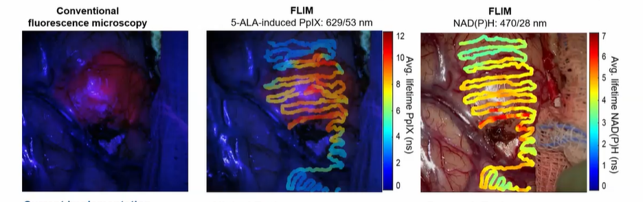
Caption: Conventional fluorescence microscopy compared to augmented reality overlays using FLIM for determining tissue types.
Image Credit: Laura Marcu
Marcu described two types of oncological surgeries where intra-operative FLIM can be used: Trans-oral robotic surgery (TORS) for head and neck cancers and neurosurgeries for primary brain cancer. Both procedures require surgeons to make visual assessments of cancerous tissues and decisions on how much tissue to excise. For head and neck cancers, the goal is to remove the tumor and a “safety margin” of healthy tissue, while brain cancer surgeries involve removing as much of the tumor as possible without damage to healthy tissues. With FLIM, surgeons can use real-time information on tissue type identification, paired with augmented reality, to delineate and visualize healthy and cancerous tissues.
Bringing FLIM to more operating rooms will require collaboration between physicists, surgeons, biologists and pathologists to develop methodologies for reconciling optical signals and gold-standard diagnostic techniques. As the technique progresses, Marcu sees FLIM as an opportunity for the optics community to make a difference in the lives of patients around the world.
30 Years of Impactful Imaging
After 30 years, OCT has become a standard of medical care, and Wolfgang Drexler explained in his plenary presentation that the best of OCT is still to come. Originally developed in 1991 by Federic Ives medal winner James Fujimoto's group at MIT, as well as pioneering work from Adolf Fercher's group in Vienna, OCT has been the fastest adopted imaging technology in the history of ophthalmology. The team had the idea to generate cross-sectional images by using local hearings interferometry methods. The technology was quickly adopted for ophthalmological applications, intravascular imaging and photodynamic therapies.
In 2015, 50 companies had footprints in OCT, mainly start-ups supported by government research. In 2022, Drexler counts 209 companies active in OCT applications, primarily in ophthalmology and endoscopy-based applications. Drexler went on to review published research on OCT, noting 74,000 thousand papers were published in 2020 on OCT-based research; an average of 21 per day. The research and industry interest is just beginning noted Drexler, as he detailed further “Big Science” investments from the National Institute of Health, National Science Foundation, and the Horizon’s 2020 Moon Program, led by Biophotonics 2022 General Chair, Rainer Andreas Leitgeb, Medizinische Universität Wien, Austria.
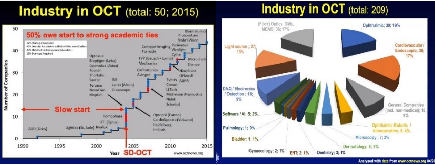
Caption: In 2015, 50 companies were active in OCT. Now, 209 companies in a number of sectors are active in OCT applications.
Image Credit: Wolfgang Drexler
What are the future growth areas in OCT? Drexler highlighted current research areas of his collaborators around the world: Intraoperative 4D OCT, Contactless Robotic OCT Scanning, Polarization Sensitive OCP for Imaging Fibrosis and OCT in a Needle Format among others. He also notes cost-effective OCT is enabled by technological advancements like photonic integration and the availability of cheaper swept-source laser technology.
Miss this year’s Biophotonics Congress? Watch the Plenary presentations on-demand: Optica Biophotonics Congress. Until next year! Biophotonics Congress: Optics in Life Sciences will take place 24 –27 April, 2023.
