New Air-Filled Fiber Bundle Could Make Endoscopes Smaller
About
24 October 2018
New Air-Filled Fiber Bundle Could Make Endoscopes Smaller
24 October 2018
New Air-Filled Fiber Bundle Could Make Endoscopes Smaller
Innovative optical fibers poised to expand endoscopy to new wavelengths and diagnoses
WASHINGTON — Researchers have fabricated a new kind of air-filled optical fiber bundle that could greatly improve endoscopes used for medical procedures like minimally invasive surgeries or bronchoscopies. The new technology might also lead to endoscopes that produce images using infrared wavelengths, which would allow diagnostic procedures that are not possible with endoscopes today.

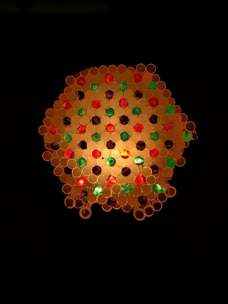
Caption: Researchers created a fiber bundle that is a honeycomb of hollow glass capillaries with solid glass cores nestled in among the glass. They did this by first making the stack of capillaries shown in the top image, with the colored ends of the solid glass cores nestled within. They heated and stretched this, making it thin enough to stack up again, producing the bottom image. This was used to make the light guiding part of the imaging fiber bundle.
Image Credit: Harry Wood, University of Bath
Endoscopes use bundles of optical fibers to transmit images from inside the body. Light falling on one end of the fiber bundle travels through each fiber to the far end, allowing a picture to be carried in the form of thousands of spots that are much like the pixels that make up a digital picture.Optical fibers consist of an inner core and an outer cladding with different optical properties, which traps the light inside and allows it to travel down the fiber. Rather than using cores and claddings made of two types of glass like most fiber bundles, the new bundles use an array of glass cores surrounded by hollow glass capillaries filled with air that act as the cladding.
In The Optical Society (OSA) journal Optics Letters, researchers show that their new fiber bundles, which they call air-clad imaging fibers, maintain the resolution of the best commercial imaging fibers at double the wavelength range that the commercial fibers can be used. The new fiber could be used to create endoscopes that are smaller or have higher resolutions than those available today.
“Higher resolution is always helpful to clinicians carrying out endoscopic procedures, but the most sensitive jobs, such as those in the brain, usually require the thinnest instruments,” said the paper’s first author, Harry Wood of the University of Bath. “These instruments are usually so narrow that the imaging fiber contains too few cores to make a clear image. Our air-clad bundles allow more fibers to be packed into a smaller diameter and so will likely be particularly useful in these situations.”
In addition to applications in medical diagnostics and treatment, the new fiber could prove useful for industrial applications such as monitoring the contents of hazardous machines or imaging the inside of oil and mineral drills.
Combining air and glass
When a bundle of fibers contains a greater number of cores within a given cross section area, it will produce more detailed images in the same way that a camera with more pixels creates higher resolution images. However, if the cores are too small and close together, light can leak from one to another and the image becomes blurry.
“The honeycomb structure we developed combines glass and air to contain light far more tightly in the cores than traditional imaging fibers that use two types of glass,” said Wood. “This allows us to bring the cores closer together than ever before possible, or squeeze in longer wavelengths of light, without the blurring that would be seen with conventional approaches.”
The fact that the new fibers work well with wavelengths further into the infrared portion of the spectrum could allow the development of endoscopes that image fluorescent markers that emit at these wavelengths. Infrared light also can be used to image cells that are embedded more deeply within tissue than can be imaged with visible wavelengths.
“There are fluorescent marker probes that emit light of specific wavelengths in response to certain bacteria or immune cells,” said Wood. “These could be very effective at highlighting disease in the lung, for example, but we can currently use only one or two such probes in the wavelength range that is offered by today’s endoscope technology.”
Comparing fiber performance
To test the imaging fibers, the researchers made an air-clad fiber bundle that matched the resolution of a leading commercial fiber because it had the same spacing between cores. They were able to incorporate more than 11,000 cores into the fiber by stacking multiple smaller honeycomb structures together.
The researchers point out that the principle behind the new fibers has been known for years but that fabrication approaches, especially for fibers with air gaps, have just recently advanced to the point where these fibers could be made.
The researchers used their new air-clad fiber bundle and the commercial fiber to image a standard test target image. “We were delighted to find that the air-clad fiber functioned well beyond the wavelength range our visible camera could detect,” said Wood. “When we changed to an infrared camera, we saw that the fiber created a clear image at double the wavelength that the commercial fiber reached.”
The researchers are now working to figure out how to seal the ends of the fibers to prevent biological material from entering the bundle and to preserve the fine glass webs between the cores. Once this problem is solved, the researchers foresee widespread adoption of this technology into next-generation endoscopes.
Caption: Tests showed that air-clad fiber bundles maintained the resolution of the best commercial imaging fibers but could be used at wavelengths into the infrared portion of the spectrum. In this image, researchers are using a white light laser to investigate the properties of their imaging fiber at a wide spectrum of wavelengths.
Image Credit: Stephanos Yerolatsitis, University of Bath
This project is part of a multi-institutional, interdisciplinary research collaboration called Proteus that brings together scientists from the Universities of Edinburgh, Heriot Watt, and Bath to improve the diagnostic technology used to guide the treatment of patients who are critically ill with lung diseases. Proteus is funded by the UK Engineering and Physical Sciences Research Council.
Paper: H. A. C. Wood, K. Harrington, T. A. Birks, J.C. Knight, J.M. Stone. “High resolution air-clad imaging fibers,” Opt. Lett., 43, 21, 5311-5314 (2018).
DOI: 10.1364/OL.43.005311.
About Optics Letters
Optics Letters offers rapid dissemination of new results in all areas of optics with short, original, peer-reviewed communications. Optics Letters covers the latest research in optical science, including optical measurements, optical components and devices, atmospheric optics, biomedical optics, Fourier optics, integrated optics, optical processing, optoelectronics, lasers, nonlinear optics, optical storage and holography, optical coherence, polarization, quantum electronics, ultrafast optical phenomena, photonic crystals and fiber optics.
About The Optical Society
Founded in 1916, The Optical Society (OSA) is the leading professional organization for scientists, engineers, students and business leaders who fuel discoveries, shape real-life applications and accelerate achievements in the science of light. Through world-renowned publications, meetings and membership initiatives, OSA provides quality research, inspired interactions and dedicated resources for its extensive global network of optics and photonics experts. For more information, visit osa.org.
Media Contact:
mediarelations@osa.org
