Head-Mounted Microscope Allows Long-Term Brain Imaging in Freely Moving Mice
About Optica
09 December 2021
Head-Mounted Microscope Allows Long-Term Brain Imaging in Freely Moving Mice
Detachable photoacoustic probe captures neurovascular dynamics that play a key role in neurological disorders
WASHINGTON — Researchers have developed the first detachable head-mounted photoacoustic microscope for imaging brain activity in freely moving mice. Because the device can be removed after imaging, it enables long-term studies that could reveal important new insights into neurodegenerative diseases and other neurological disorders.
“Epilepsy, Alzheimer's disease and Parkinson's disease can all seriously interrupt neurovascular coupling—the link between neural activity and subsequent changes in cerebral blood flow,” said research team leader Lei Xi from the Southern University of Science and Technology in China. “Our new probe is ideal for studying neurovascular coupling because it has the potential to capture the dynamics of both neuron and vascular networks simultaneously.”
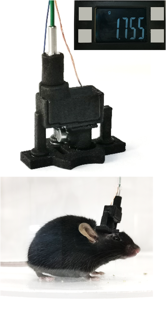
Caption: Researchers have developed a head-mounted photoacoustic microscope that can image the brain activity in freely moving mice. The probe can be removed after imaging, making long-term studies possible.
Credit: Lei Xi, Southern University of Science and Technology
The new microscope probe weighs just 1.8 grams and is based on optical resolution photoacoustic microscopy (ORPAM), which can capture anatomical and functional dynamics of the brain without requiring the use of fluorescent tags or labels. In the Optica Publishing Group journal Optics Letters, Xi and colleagues describe how they optimized the design of the new probe to make it light enough for use in freely moving mice.
“Head-mounted microscopes that use multi-photon or fluorescence imaging primarily capture the activities of single neurons,” said Xi. “Our ORPAM probe can capture cerebral vascular network and hemodynamics of large portions of the cerebral cortex with capillary-level resolution without requiring any labels.”
Miniaturizing a microscope
The new work builds on a wearable ORPAM probe the researchers previously built for freely moving rats. Although it performed well, it had to be permanently fixed on the rat and was too large and heavy to be carried around by mice, which are the preferred animal models for many brain studies.
To make a smaller probe, the researchers used optical simulation calculations to carefully optimize the entire light path of the microscope. They also selected high-performance miniature components including an aspheric lens, micro-electro-mechanical-system (MEMS) scanner and customized miniaturized piezoelectric ultrasonic detector.
The resulting probe weighs less than 10% of an adult mouse’s bodyweight, has a resolution of 2.8 microns and can image a large field of view of 3×3 mm2. To make it detachable, the researchers incorporated three pairs of magnets that connect the imaging probe with a lightweight mounting base attached to the mouse’s skull. The probe can be removed directly after imaging and then reinstalled later, enabling repeated, long-term monitoring of freely moving animals.
Long-term imaging
After evaluating the probe’s performance on a synthetic material that mimics soft tissue, the researchers used it to image vascular networks in the cerebral cortex of a mouse for 40 minutes. They also conducted long-term monitoring experiments that lasted for seven days. The results from these tests showed that the probe can provide stable and high-quality ORPAM images in freely moving mice.
“In the future, we intend to develop a probe with capillary-level resolution, video-rate imaging speed and a field of view large enough to capture the entire mouse cerebral cortex,” said Xi. “We want to push the performance of the probe so that even more can be learned about brain activity and how it might relate to disease and health.”
Paper: H. Guo, Q. Chen, W. Qin, W. Qi, L. Xi, “Detachable head-mounted photoacoustic microscope in freely moving mice,” Opt. Lett., 46, 6055-6058 (2021).
DOI: https://doi.org/10.1364/OL.444226
About Optica Publishing Group
Optica Publishing Group is a division of the society, Optica, Advancing Optics and Photonics Worldwide. It publishes the largest collection of peer-reviewed and most-cited content in optics and photonics, including 19 prestigious journals, the society’s flagship member magazine, and papers and videos from over 1200 conferences. With over 520,000 journal articles, conference papers and videos to search, discover and access, its publications portfolio represents the full range of research in the field from around the globe.
About Optics Letters
Optics Letters offers rapid dissemination of new results in all areas of optical science with short, original, peer-reviewed communications. Optics Letters accepts papers that are noteworthy to a substantial part of the optics community. Published by Optica Publishing Group and led by Editor-in-Chief Carsten Rockstuhl, Karlsruher Institut für Technologie, Germany. For more information, visit Optics Letters.
Media Contact
