Noninvasive stent imaging powered by light and sound
About Optica
25 July 2025
Noninvasive stent imaging powered by light and sound
Photoacoustic microscopy shows promise for monitoring stents through the skin, without surgery or radiation
WASHINGTON — In a new study, researchers show, for the first time, that photoacoustic microscopy can image stents through skin, potentially offering a safer, easier way to monitor these life-saving devices. Each year, around 2 million people in the U.S. are implanted with a stent to improve blood flow in narrowed or blocked arteries.
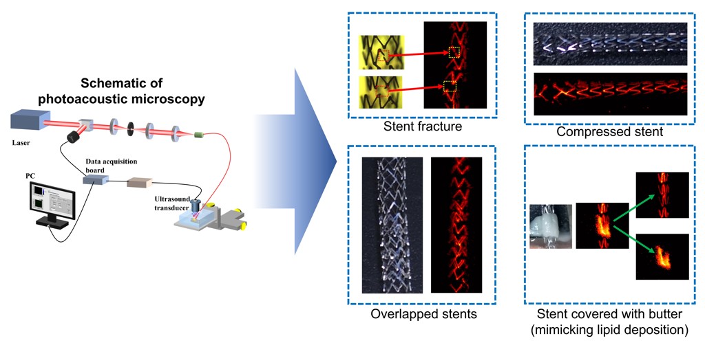
Caption: Researchers used photoacoustic microscopy to image stents through skin. They were able to visualize stents with fractures and compression and other clinical scenarios such as overlapped stents or conditions mimicking lipid deposition.
Credit: Myeongsu Seong, Xi’an Jiaotong-Liverpool University“It is critical to monitor stents for problems such as fractures or improper positioning, but conventionally used techniques require invasive procedures or radiation exposure,” said co-lead researcher Myeongsu Seong from Xi’an Jiaotong-Liverpool University in China. “This inspired us to test the potential of using photoacoustic imaging for monitoring stents through the skin.”
In the Optica Publishing Group journal Optics Letters, the researchers show that photoacoustic microscopy can be used to visualize stents covered with mouse skin under various clinically relevant conditions, including simulated damage and plaque buildup.
“While our photoacoustic microscopy results are preliminary, further development could enable frequent, noninvasive monitoring of stent status — without the need for surgical access or X-ray exposure,” said co-lead researcher Sung-Liang Chen from Shanghai Jiao Tong University in China. “This would make it easier and safer to monitor the condition of stents in patients.”
Using sound to see stents
Photoacoustic imaging is a label-free technique that detects sound waves generated when materials absorb light and release energy. Because sound scatters less than light, this imaging method can be used to acquire higher-resolution images at greater depths than purely optical methods.
Although other studies have used photoacoustic imaging via an endoscope to image stents, this still requires that patients undergo a procedure. In the new study, the researchers examined whether photoacoustic microscopy might enable noninvasive stent monitoring through the skin.
To do this, they mimicked different stent scenarios, including fractures, compression and movement of overlapped stents. They also used butter to mimic deposition of plaque or blood clots after stenting. Using photoacoustic microscopy at various wavelengths, including 670 nm and 1210 nm, they were able to image these various stent conditions through excised mouse skin.
“One of the most interesting results is that we could easily differentiate between the butter we used to mimic a lipid plaque and the stent,” said Seong. “Because plaque and stents absorb light differently, using two wavelengths helped us distinguish them.”
Adapting for depth
The researchers say that photoacoustic microscopy could potentially be used to image stents placed in dialysis access sites, which are typically located just beneath the skin. For stents in deeper areas like the carotid artery, a related method called photoacoustic computed tomography may be more suitable.
The researchers point out that before photoacoustic imaging can be used for clinical noninvasive stent monitoring, in vivo animal experiments and preliminary clinical experiments would have to be performed. The system would also need to be optimized for use in various parts of the body.
Paper: S. Liang, N. Wan, W. Pi, K. Zhang, J. Wen, S. Chen, M. Seong, “Photoacoustic microscopy for visualization of stents in multiple scenarios,” Opt. Lett., 50, 4722-4725 (2025).
DOI: 10.1364/OL.564778
About Optica Publishing Group
Optica Publishing Group is a division of the society, Optica, Advancing Optics and Photonics Worldwide. It publishes the largest collection of peer-reviewed and most-cited content in optics and photonics, including 19 prestigious journals, the society’s flagship member magazine, and papers and videos from over 1200 conferences. With over 505,000 journal articles, conference papers and videos to search, discover and access, its publications portfolio represents the full range of research in the field from around the globe.
About Optics Letters
Optics Letters offers rapid dissemination of new results in all areas of optical science with short, original, peer-reviewed communications. Optics Letters accepts papers that are noteworthy to a substantial part of the optics community. Published by Optica Publishing Group and led by Editor-in-Chief Carsten Rockstuhl, Karlsruher Institut für Technologie, Germany. For more information, visit Optics Letters.
Media Contact
