About
26 March 2024
Structured illumination microscopy shows promise for detecting prostate cancer margins
Results from clinical study indicate SIM is fast and accurate enough for use during surgery
Results from a new clinical study demonstrate the potential of structured illumination microscopy (SIM) for detecting prostate cancer margins during surgery. This capability could help surgeons ensure that all the cancerous tissue is removed, which would reduce cancer recurrence and improve the chances of successful treatment.
SIM is a wide-field optical sectioning fluorescence microscopy technique that projects patterned light onto a sample. The resulting interference patterns are used to reconstruct images with higher resolution than traditional microscopy methods. Compared to similar techniques, SIM offers faster surface imaging, making it particularly well-suited for capturing high-resolution images of large tissue samples in nearly real-time.
The new study shows that SIM not just feasible but also fast and accurate enough for intraoperative tumor margin imaging. Ivan Bozic from Tulane University will detail the work at Optica’s Biophotonics Congress being held in Fort Lauderdale, Florida, 07 – 10 April 2024. Bozic’s presentation is scheduled for Tuesday, 10 April at 8:00-8:15 EDT.
“Most of the time, when surgeons remove a cancerous tumor, they are doing so blind to the presence of cancer cells at the edge of the tumor, since these cells are microscopic and too small to see by the naked eye,” said Bozic. As a result, sometimes tumor is left inside the patient after surgery, and this is not known until days or weeks later, when is too late to correct the surgery, leaving the possibility that the remaining tumor can grow. If the tumor edge could be scanned at a microscopic level in real-time during the surgery, then surgeons could identify and remove any residual tumor from the patient, improving outcomes and prognosis for the patient."
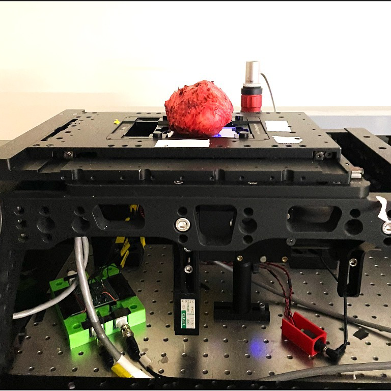
For the new research, the investigators used SIM to acquire surface images of 172 prostates that had been removed for cancer treatment. The microscopy system had a resolution of 1.3 microns and a single-frame field of view of 1.3 mm x 1.3 mm. The researchers performed image reconstruction and coloring with a square law detection reconstruction algorithm and an analytical false coloring approach. Large image panels were stitched together.
They reported multi-scale imaging could be performed at speeds of 1 cm2 per minute or faster. This enabled an entire prostate surface — over 10 gigapixels of data covering up to 60 square centimeters per patient — to be imaged in less than 1 hour.
The researchers were able to detect histologically confirmed positive surgical margins – ones where cancer cells were still present — on the clinical margin surface using the SIM method. They also showed that the approach could identify positive surgical margins that were not visible using traditional histological analysis.
The researchers are now analyzing patient outcomes to verify the accuracy of their findings.
“Our work has demonstrated the feasibility and potential clinical utility of the SIM imaging approach for scanning tumor surfaces during surgery. Our current and future work is aimed at improving the speed and automation of the device to make it practical for widespread routine clinical use in the operating room,” said Bozic.
About Optica Biophotonics Congress
Optica Biophotonics Congress is an annual meeting that focuses on biomedical optics and optics in the life sciences in alternating years. The 2024 meeting, Optica Biophotonics Congress: Biomedical Optics, Biomedical Optics focuses on technological solutions to medical challenges and medical applications. The meeting will be held as an in-person event with on-demand content. More information at https://www.optica.org/events/congress/biophotonics_congress_biomedical_optics/.
About Optica
Optica, Advancing Optics and Photonics Worldwide, is the society dedicated to promoting the generation, application, archiving and dissemination of knowledge in the field. Founded in 1916, it is the leading organization for scientists, engineers, business professionals, students and others interested in the science of light. Optica's renowned publications, meetings, online resources and in-person activities fuel discoveries, shape real-life applications and accelerate scientific, technical and educational achievement. Discover more at: Optica.org
Media Contact
