Researchers add swept illumination to open-top light-sheet microscope
About Optica
20 March 2024
Researchers add swept illumination to open-top light-sheet microscope
Advance improves field of view and optical sectioning, laying groundwork for 3D pathology applications
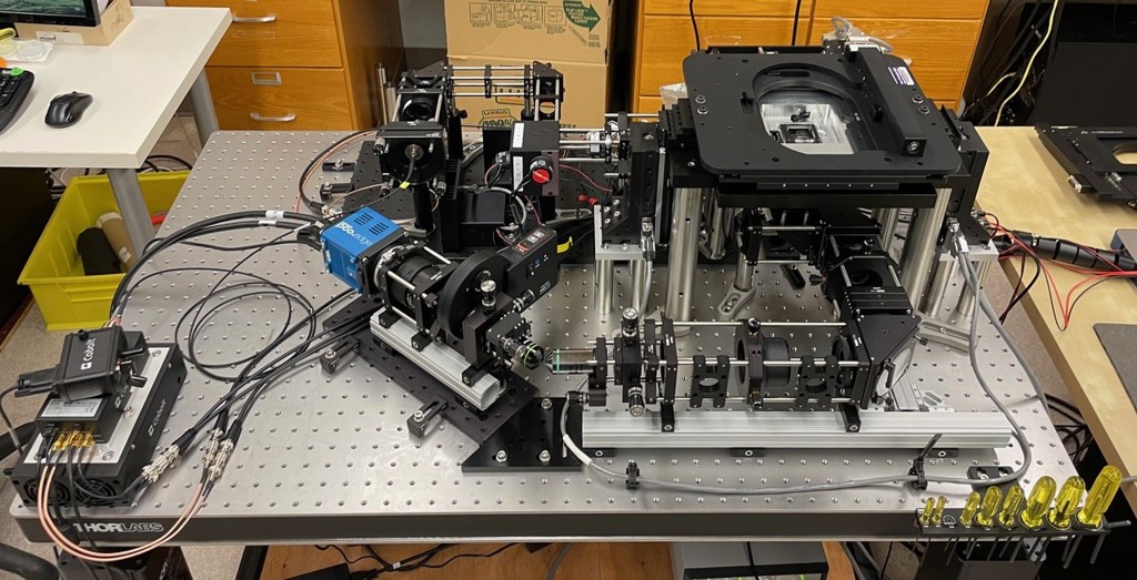
Researchers have incorporated a swept illumination source into an open-top light-sheet microscope to enable improved optical sectioning over a larger area of view. The advance makes the technique more practical for nondestructive 3D pathology.
3D pathology is being explored as an alternative to traditional slide-based histology because it can provide detailed 3D insights into pathological structures and cellular interactions without altering the tissue. This approach makes it possible to analyze complex 3D tissue structures and to image thick tissues, which is not possible with slide-based methods.
The researchers used their improved open-top light-sheet microscope to capture images of densely labeled clinical specimens, showing its potential for nondestructive 3D pathology. Kevin W. Bishop from the University of Washington will detail the work at Optica’s Biophotonics Congress being held in Fort Lauderdale, Florida, 07 – 10 April 2024. Bishop’s presentation is scheduled for Tuesday, 09 April at 13:45 - 14:00 EDT.
"For certain diseases, like prostate cancer, it can be challenging to determine which patients need aggressive treatment and which patients do not. 3D information could ultimately help clinicians better determine the best course of treatment for each patient," said Bishop.
Open-top light-sheet microscopy is used to rapidly acquire 3D images of fluorescently labeled tissues that have been treated in a way that makes them transparent or translucent. The typical setup uses a fixed thin sheet of light to illuminate and image the sample from below, much like a flatbed document scanner. This enables high-resolution imaging of large areas at much faster speeds than are possible with other 3D imaging approaches (e.g. confocal microscopy).
Although many types of labels can be used with this microscopy technique, 3D pathology samples typically use dyes that mimic the hematoxylin and eosin (H&E) staining used in traditional histology slides. Because this type of staining is much denser than highly targeted stains, the microscope’s optical-sectioning capability — its ability to visualize a thin slice within a 3D sample — becomes key to achieving good image quality.
Although better sectioning is possible by using a higher illumination numerical aperture, this creates a shorter depth of focus that reduces the system’s usable field of view. To overcome this challenge, the researchers developed a new open-top light-sheet microscope that incorporates an axially swept illumination arm.
Compared to their previous microscope design with a fixed light sheet, the new system quadrupled the field of view and doubled the optical sectioning ability without compromising volumetric imaging speed. The researchers demonstrated its usefulness by imaging a densely labeled cleared mouse kidney. They also acquired other datasets from clinical tissues to further show that the optimized system can deliver the image quality and field of view necessary for 3D pathology studies.
“We plan to use this platform to run large-scale clinical studies that will help us understand where 3D pathology can have the greatest clinical impact,” said Bishop.
About Optica Biophotonics Congress
Optica Biophotonics Congress is an annual meeting that focuses on biomedical optics and optics in the life sciences in alternating years. The 2024 meeting, Optica Biophotonics Congress: Biomedical Optics, Biomedical Optics focuses on technological solutions to medical challenges and medical applications. The meeting will be held as an in-person event with on-demand content. More information at https://www.optica.org/events/congress/biophotonics_congress_biomedical_optics/.
About Optica
Optica, Advancing Optics and Photonics Worldwide, is the Society dedicated to promoting the generation, application, archiving and dissemination of knowledge in the field. Founded in 1916, it is the leading organization for scientists, engineers, business professionals, students and others interested in the science of light. Optica's renowned publications, meetings, online resources and in-person activities fuel discoveries, shape real-life applications and accelerate scientific, technical and educational achievement. Discover more at: Optica.org
Media Contact
