X-ray method enables micron-resolution imaging of living organisms over long time periods
About Optica
07 December 2023
X-ray method enables micron-resolution imaging of living organisms over long time periods
By lowering the X-ray dose needed for imaging, new approach extends possibilities for biological and biomedical research
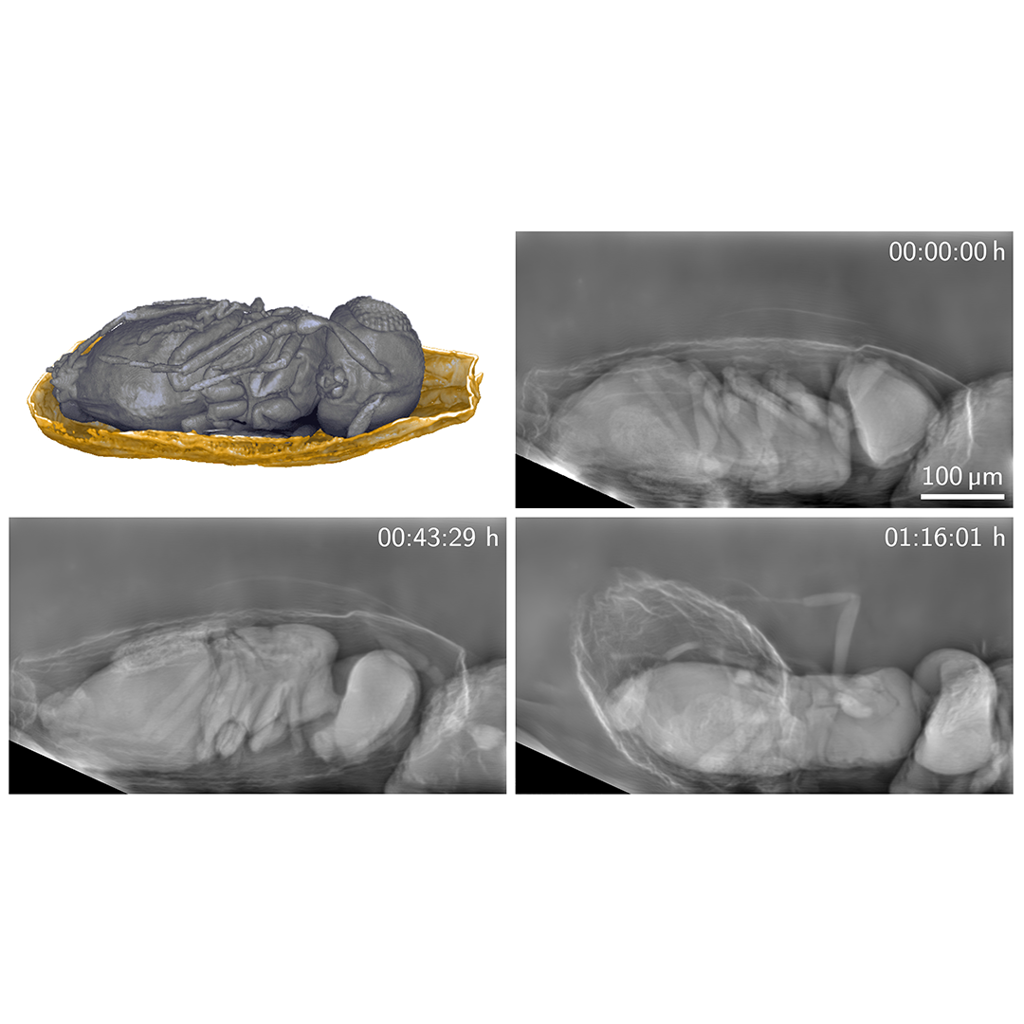
Caption: A new X-ray imaging technique can produce detailed images of living organisms with a much lower X-ray dose than previously possible. The researchers used the new technique to image tiny parasitoid wasps emerging from their host eggs for more than 30 minutes.
Credit: Rebecca Spiecker, Karlsruhe Institute of Technology
WASHINGTON — Researchers have developed an X-ray imaging technique that can produce detailed images of living organisms with a much lower X-ray dose than previously possible. The advance enables small organisms or other sensitive samples to be studied at high resolution over much longer periods, which could reveal new insights into a variety of dynamic processes.
The approach is based on phase contrast imaging, which relies not only on the absorption of X-rays in a sample, but also on the wave properties of X-rays. More precisely, it creates images from phase changes that occur as X-rays go through a specimen. “Previously, micrometer resolution X-ray phase contrast imaging of living organisms was only possible for a few seconds up to minutes because severe radiation damage would occur,” explained researcher team member Rebecca Spiecker from Karlsruhe Institute of Technology in Germany. “We reduced the necessary X-ray dose by overcoming the current limitations of high-resolution imaging for dose-sensitive applications.”
In Optica, Optica Publishing Group’s journal for high-impact research, the researchers describe how they developed a new X-ray imaging system that uses dedicated highly-efficient X-ray optics and single-photon-counting detectors to boost the dose efficiency for full-field imaging at micrometer resolution. They demonstrated the benefit of the new technique by imaging tiny parasitoid wasps emerging from their host eggs for more than 30 minutes.
“We show that our method exhibits superior imaging performance compared to a conventional high-resolution detector,” said Spiecker. “This could be useful, for example, for capturing details of the development and behavior of small model organisms, such as Xenopus frog embryos, over a longer time scale than is currently possible.”
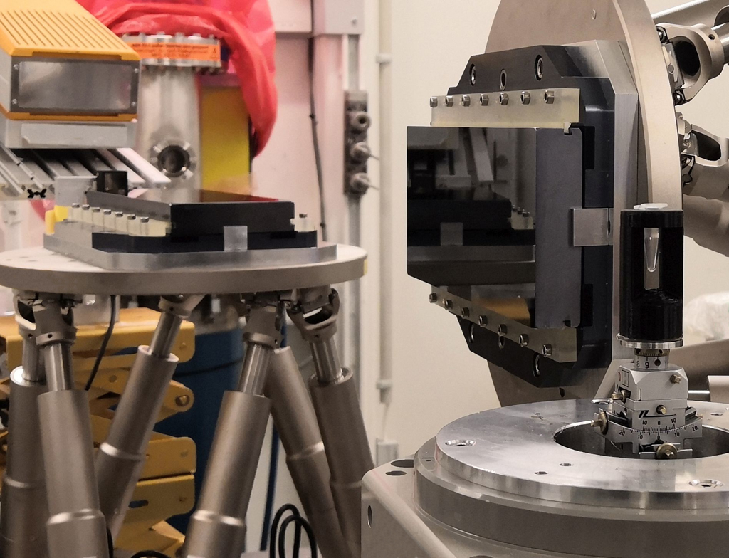
Caption: The new X-ray imaging technique uses a much lower X-ray dose thanks to two Bragg magnifier crystals (center) and a single-photon-counting detector (on the left). The sample is shown on the right.
Credit: Rebecca Spiecker, Karlsruhe Institute of Technology
Better images with less radiation
X-ray imaging can reveal hidden structures and processes in living organisms. However, it also exposes organisms to radiation that is harmful at high doses, limiting how long observations can last before damage occurs. This is aggravated by the fact that the detection efficiency of commonly used high-resolution detectors decreases with increasing resolution, which means that even higher X-ray doses are necessary to obtain a high-resolution image.
To overcome this challenge, the researchers developed a phase contrast imaging approach that directly magnifies the X-ray image rather than converting the X-ray image into a visible light image and then magnifying it, which is the typical method. This allowed them to use highly efficient large-area detectors while maintaining micrometer spatial resolution.
In the new imaging system, the researchers used a single-photon-counting imaging detector with a pixel size of 55 microns. The X-ray image is magnified behind the sample using crystal optics, known as a Bragg magnifier. The latter consists of two perfect silicon crystals to perform magnification.
“To achieve the highest possible dose efficiency for full-field X-ray imaging at micrometer resolution, we combine X-ray phase contrast, a Bragg magnifier and a single-photon-counting detector, all optimized for an optimal X-ray energy of 30 keV,” said Spiecker. “The concept of Bragg magnifiers dates back to the late 1970s, and although their potential for increasing dose efficiency has been noted, it has not been explored until now.”
After showing that their new system could attain a dose efficiency of more than 90% while providing a resolution of up to 1.3 microns, the researchers compared its performance to a conventional high-resolution detector system using the same sample, X-ray fluence and 30 keV X-ray energy. “At this energy, we showed that the detective quantum efficiency of our system exceeds the conventional system by over two orders of magnitude for the relevant high-resolution components of the image,” said Spiecker. “This results in better images and allows a drastic reduction in the X-ray dose in the sample.”
Caption: The researchers used the new technique to image tiny parasitoid wasps emerging from their host eggs. Even after 30 minutes of imaging, the wasps did not show any abnormalities in their behavior thanks to the minimal radiation exposure.
Credit: Rebecca Spiecker, Karlsruhe Institute of Technology
Imaging tiny insects
The researchers then used the system to perform a pilot behavioral study on living parasitoid wasps, which are widely used for biological pest control. Thanks to the minimal radiation exposure, they were able to capture images of the tiny wasps inside their host eggs for 30 minutes before the wasps finally emerged.
The researchers say that the method might also be useful for biomedical applications, such as gentle tomographic examination of biopsy samples. However, using a Bragg magnifier requires a monochromatic, coherent and collimated beam, which is available at X-ray synchrotron facilities. They are also continuing to improve the system to achieve a larger field of view and increased long-term mechanical stability for even longer measurement times.
Paper: R. Spiecker, P. Pfeiffer, A. Biswal, M. Shcherbinin, M. Spiecker, H. Hessdorfer, M. Hurst, Y. Zharov, V. Bellucci, T. Farago, M. Zuber, A. Herz, A. Cecilia, M. Czyzycki, C. S. Baraldi Dias, D. Novikov, L. Krogmann, E. Hamann, T. van de Kamp, T. Baumbach, “Dose-efficient in vivo X-ray phase contrast imaging at micrometer resolution by Bragg magnifiers,” 10, 12, 1633-1640 (2023).
DOI: doi.org/10.1364/OPTICA.500978
About Optica Publishing Group
Optica Publishing Group is a division of the society, Optica, Advancing Optics and Photonics Worldwide. It publishes the largest collection of peer-reviewed and most-cited content in optics and photonics, including 19 prestigious journals, the society’s flagship member magazine, and papers and videos from over 1200 conferences. With over 520,000 journal articles, conference papers and videos to search, discover and access, its publications portfolio represents the full range of research in the field from around the globe.
About Optica
Optica is an open-access journal dedicated to the rapid dissemination of high-impact peer-reviewed research across the entire spectrum of optics and photonics. Published monthly by Optica Publishing Group, the Journal provides a forum for pioneering research to be swiftly accessed by the international community, whether that research is theoretical or experimental, fundamental or applied. Optica maintains a distinguished editorial board of more than 60 associate editors from around the world and is overseen by Editor-in-Chief Thomas Krauss, University of York, UK. For more information, visit Optica.
Media Contact
