21 - 24 April, 2025
Loews Coronado Bay Resort
Coronado, California USA
Bio-Optics: Design and Applications
Novel Techniques in Microscopy
Optical Manipulation and Its Applications
Optical Molecular Probes, Imaging and Drug Delivery
Optics and the Brain
Novel Techniques in Microscopy
Optical Manipulation and Its Applications
Optical Molecular Probes, Imaging and Drug Delivery
Optics and the Brain
Bio-Optics: Design and Applications
-
Hatice Ceylan Koydemir, Texas A&M University, United States
Smart and Portable Technologies Engineered for Biomedical Sensing and Imaging -
Arnaud Dubois, Institut d'Optique Graduate School, France
Line-Field Confocal Optical Coherence Tomography (LC-OCT) for Dermatology -
Pilhan Kim, Korea Advanced Inst of Science & Tech, Republic Of Korea
Real-Time Intravital Microscopy with Suction-Assisted Imaging Window for Thoracic Organs -
Pierre Lane, Simon Fraser University, Canada
Endoscopic OCT-AFI – In Vivo Imaging of the Lungs, Oral Cavity, and Gynecologic Tract -
Muyinatu Lediju Bell, Johns Hopkins University, United States
Listening to the Sound of Light to Guide Surgeries -
Jonathan Liu, University of Washington, United States
Non-Destructive 3D Pathology and Analysis for Precision Medicine -
Andrea Locke, Vanderbilt University, United States
Raman-Based Spectroscopic Tools for Characterizing Pediatric Eosinophilic Esophagitis -
Christine O'Brien, Washington University School of Medicine, United States
Light-Based Detection of Postpartum Hemorrhage with a Wearable Device -
Juergen Popp, Friedrich-Schiller-Universität Jena, Germany
Multimodal Imaging Together with AI – A Powerful Toolbox for Tumor Diagnosis -
Marinko Sarunic, Simon Fraser University, United Kingdom
Deep Learning Methods for Sensorless Adaptive Optics -
Serhat Tozburun, Izmir Biomedicine and Genome Center, Turkey
Guide Mapping and Endoscopy Cap Approaches for Well-Confined Mucosal Laser Coagulation -
Néstor Uribe-Patarroyo, Massachusetts General Hospital, United States
Probabilistic Structural and Functional Optical Coherence Tomography
Novel Techniques in Microscopy
-
Adela Ben-Yakar, University of Texas at Austin, United States
LEAD Fluorescence Microscopy Performing at 0.8 Million Frames Per Second for 3D Imaging Flow Cytometry and Brain Imaging -
Bo-Jui Chang, UT Southwestern Medical Center at Dallas, United States
Pushing the Limits with Structured Illumination in Light-Sheet Fluorescence Microscopy -
Vincent Ralph Ching-Roa, University of Rochester, United States
Rapid Large-Volume Tissue Imaging with Multiplane Parallel Strip-Scanning Two-Photon Microscopy -
Wonshik Choi, Korea University, Republic Of Korea
Solving Higher Order Inverse Scattering Problem for In Vivo Through-Skull Imaging -
Keng Chou, University of British Columbia, Canada
Spinning-Disk Structured Illumination Microscopy for High-Speed Super-Resolution Imaging -
Shwetadwip Chowdhury, University of Texas at Austin, United States
3D Tomography of Scattering Tissue -
Michelle Digman, University of California Irvine, United States
The Phasor Approach to FLIM for Metabolic Profiling and Mitometer for Tracking Phenotypic Changes in Mitochondria in Cancer Cells -
Adam Glaser, Allen Institute for Neural Dynamics, United States
Leveraging Electronics Metrology Technologies for a New Large-Scale Light-Sheet Microscopy System -
Sicong He, Southern Univ of Science & Technology, China
Minimally Invasive Imaging of the Mouse Spinal Cord at Subcellular Resolution Through an Optically Cleared Intervertebral Window -
Guangwei Hu, Nanyang Technological University, Singapore
Single-Shot, Isotropic and Miniaturized Differential Interference Contrast (SIM-DIC) Microscopy Based on Computational Flat-Optics -
Fang Huang, Purdue University, United States
In Situ PSF Retrieval in 4Pi Single-Molecule Systems Enables Ultra-High-Resolution Imaging Through Brain Slices With Uncompromised Fidelity -
Zhiwei Huang, National University of Singapore, Singapore
Scan-Free Stimulated Raman Scattering Tomography for Volumetric Deeper Tissue Imaging -
Xingchen Ji, Shanghai Jiao Tong University, China
Developing High Confinement and Low-Loss Silicon Nitride Photonic Devices for Optical Coherence Tomography -
Xingde Li, Johns Hopkins University, United States
Two-Photon Fiberscopy Imaging of Neural Activities in Freely-Behaving Rodents and Beyond -
Chang Liu, Helmholtz Institute Jena, Germany
Revealing the Ultra-Structure of Microorganisms Using Table-Top Extreme Ultraviolet Ptychography -
Kevin Tsia, University of Hong Kong, Hong Kong
Speeding in Microscopy: from Optofluidic Single-Cell Imaging to In-vivo Animal Imaging -
Tom Vettenburg, University of Dundee, United Kingdom
Planar Light-sheet Microscopy with Curved Airy Beams -
Dushan Wadduwage, Harvard University, United States
Differentiable Microscopy (∂μ) as a Generalized Paradigm of Optics and Optical System Design -
Yuxiao Wei, Harvard Medical School, United States
Quantitative Analysis of Drug Tablet Aging by Fast Hyperspectral Stimulated Raman Scattering Microscopy -
Florian Willomitzer, Univ of Arizona, Coll of Opt Sciences, United States
Macroscopic Imaging through Scattering Media with Single-Shot Synthetic Wavelength Holography -
Sixian You, Massachusetts Institute of Technology, United States
An Energetic, Tunable, Ultra-Broadband Fiber Source for Imaging and Spectroscopy -
Roger Zemp, University of Alberta, Canada
Photoacoustic Remote Sensing Virtual Histology -
Haishan Zeng, BC Cancer Agency Research Centre, Canada
In vivo Multiphoton Microscopy and Multiphoton Absorption Based Laser Therapy -
Alessandro Zunino, Istituto Italiano di Tecnologia, Italy
Extending the Three-Dimensional Resolution with Focus-ISM
Optical Manipulation and Its Applications
-
John Bechhoefer, Simon Fraser University, Canada
Information Engines Based on Optical Traps and Feedback -
Pierre-Francois Brevet, Université de Lyon, France
Second Harmonic Generation from Nanoparticles : From Free Liquid Suspensions to Controlled Single Nanoparticles -
Grégory David, ETH Zurich, Switzerland
Optical Trapping of Aerosols -
Anna Chiara De Luca, CNR ISASI, Italy
SERS-Based Biosensors for Biomedical Applications -
Jochen Fick, Institut Néel, France
Optical Fiber Tweezers for Biological Specimen Trapping -
Nancy Forde, Simon Fraser University, Canada
Manipulation beyond Light: Using a Centrifuge for High-Throughput Single-Molecule Force Spectroscopy -
Reuven Gordon, University of Victoria, Canada
Analysis of Single Unmodified Proteins and Their Interactions with Nanoaperture Optical Tweezers: PR65 Case Study -
Patricia Haro Gonzalez, Universidad Autonoma de Madrid, Spain
Multiparametric Remote Optical Sensing by a Single Trap Upconverting Microparticle at the Microscale -
Akihiro Kusumi, Okinawa Inst of Science & Technology, Japan
Development of Ultrafast Camera-Based Single Fluorescent-Molecule Imaging for Cell Biology -
Alessandro Magazzù, CNR IPCF, Italy
Optical and Raman Tweezers for the Manipulation and Characterization of Cosmic Dust and Sea Microplastics -
Yoko Miyamoto, University of Electro-Communications, Japan
Vortex Inversion and Angular Momentum Distribution -
Justus Ndukaife, Vanderbilt University,
Trapping and Sensing Nanoscale Extracellular Vesicles and Particles Using Resonant Dielectric Cavities -
Peter Pauzauskie, University of Washington, United States
Cold Brownian Biophotonics with Nanoscale Materials -
Basudev Roy, Indian Institute of Technology Madras, India
Study of Out-of-Plane Rotations in Optical Tweezers with Usage in Soft Matter and Biological Systems -
Stephen Simpson, Institute of Scientific Instruments ASCR, Czech Republic
Non-Conservative Levitational Optomechanics: From Stochastic Bifurcations to Synchronization -
Giuseppe Strangi , Case Western Reserve University, United States
Hyperbolic Metamaterials: From Biosensing to Optomechanics -
Grover Swartzlander, Rochester Institute of Technology, United States
Applications of Radiation Pressure Force on a Thin Diffractive Film -
Fan Wang, Beihang University, China
Lanthanide Ion Modulated Dielectric Resonance Enhancement for Nanoscale Optical Force Dye and Interferometric Scattering Microscopy -
Cuifeng Ying, Nottingham Trent University, United Kingdom
Plasmonic Nanotweezers for Monitoring Conformational Dynamics of Single, Unmodified Proteins
Optical Molecular Probes, Imaging and Drug Delivery
-
Stefan Andersson Engels, Tyndall National Institute, Ireland
Upconverting Nanoparticles for High-Contrast Breast Cancer Immune-Histochemistry Biomarker Mapping -
Ali Azhdarinia, UT Health Science Center at Houston, United States
Fluorescence-Guided Surgery in Neuroendocrine Tumors: Pathway to Translation -
Carolyn Bayer, Tulane University, United States
Photoacoustic Imaging of Systemic Vasodilation -
Françoise Cailler, SurgiMab, France
Development and Clinical Application of SGM-101, a Fluorescent Anti-CEA Antibody, for Real-Time Detection of Tumors During Fluorescence-Guided Surgery -
Ji-Xin Cheng, Boston University, United States
Bond-Selective Imaging at the Same Sensitivity and Resolution of Fluorescence Microscopy -
Hak Soo Choi, Massachusetts General Hospital, United States
How to Improve Signal-to-Background Ratio in Cancer Theranosis? -
Paula Demétrio de Souza França, Memorial Sloan Kettering Cancer Center, United States
PARPi-FL as a Tool for Fluorescence-Guided Diagnosis and Intraoperative Assessment of Surgical Margins in Oral Cancer -
Summer Gibbs, Oregon Health and Science University, United States
Near Infrared Contrast Agents for Intraoperative Nerve Visualization -
Christa Haase, Massachusetts General Hospital, United States
Image-Guided Single Cell Transcriptional Analysis for Uncovering Novel Therapeutic Targets -
Tayyaba Hasan, Massachusetts General Hospital, United States
Image Guided Therapuetics -
Susanne Kossatz, Technische Universität Munchen, Germany
Fluorescent Analogs of Clinical Stage SSTR2-targeted Radioligand Therapy Agents Provide New Insights into Mechanisms of Hematotoxicity -
Twan Lammers, Rheinish Westfalische Tech Hoch Aachen, Germany
Theranostic Concepts in Cancer Nanomedicine -
Christie Lin, OnLume Surgical, United States
Real-Time Surgical Assessment Using an Ambient-Light Compatible Wide-Field Fluorescence-Guided Surgery Platform -
Guolan Lu, Stanford University, United States
Tracking Therapeutic Antibody Delivery in Clinical Tumors -
Geoffrey Luke, Dartmouth College, United States
Perfluorocarbon Nanodroplets as Multimodal Contrast Agents and Drug Delivery Vehicles -
Srivalleesha Mallidi, Tufts University, United States
Photoacoustic Imaging -
Fay Nicolson, Dana-Farber Cancer Institute, United States
Surface Enhanced Spatially Offset Raman Spectroscopy (SESORS) for Preclinical Cancer Imaging -
Girgis Obaid, University of Texas at Dallas, United States
Molecularly Targeted PDT Agents For Tumor Priming and Surgical Navigation -
Miguel Pleitez, Technische Universität Munchen, Germany
Label-Free Metabolic Microscopy by Mid-Infrared Optoacoustic and Optothermal Detection -
Narasimhan Rajaram, University of Arkansas, United States
Imaging and Sensing of Biomolecular Processes and Pathways in Primary Tumors to Identify Long-Term Outcome -
MOHAMMAD SAAD, Massachusetts General Hospital, United States
Evaluating the Imaging and Therapeutic Performance of a Dual Function Antibody Conjugate in Head and Neck Cancer Spheroids -
Kimberley Samkoe, Dartmouth College, United States
Fluorescence Guided Surgery -
Kenneth Tichauer, Illinois Institute of Technology, United States
Clinical Fluorescein Imaging to Detect Early Signs of Diabetic Retinopathy -
Danielle Tokarz, Saint Mary's University, Canada
Measurement of the Crystalline Structure of Collagen-Like Scaffolds of Otoconia in the Mouse Vestibular System by Second Harmonic Generation Microscopy
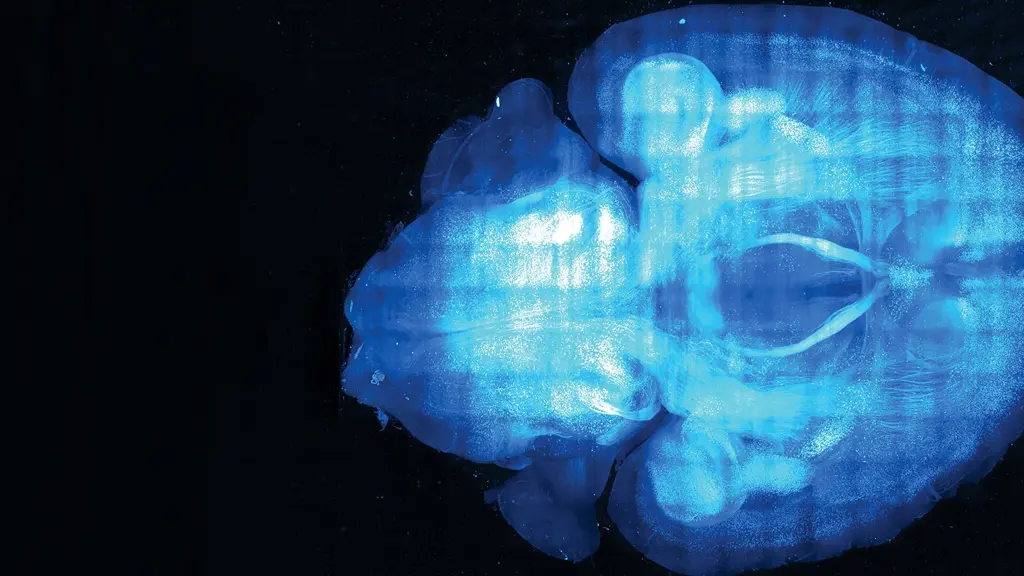
Optics and the Brain
-
Daniel Aharoni, University of California Los Angeles, United States
New Tools for Imaging Large-Scale Network Dynamics in Behaving Animals -
Sage Aronson, Neurophotometrics LLC, United States
Imaging Modality; Fiber Photometry -
Kevin Briggman, MPI Neurobiologie des Verhaltens -CAESAR, Germany
High Throughput Electron Microscopy-Based Connectomics -
Jerry Chen, Boston University, United States
Imaging the Mammalian Brain Across Space and Time with Multi-Foci Multi-Photon Microscopy -
Yao Chen, Washington University in St Louis, United States
Illuminating Neuromodulator Signaling with Fluorescence Lifetime Imaging Microscopy -
Anna Devor, Boston University, United States
Multimodal Imaging -
Dan Dombeck, Northwestern University, United States
Studying the Mouse Navigation System Using Virtual Reality and Functional Microscopy -
Elizabeth Hillman, Columbia University, United States
High Speed Volumetric Imaging -
Jung Ho Hyun, Daegu Gyeongbuk Inst of Science and Tech, United States
Tagging and Manipulating Neural Ensembles to Help Treat Brain Illness -
Alexandr Klioutchnikov, MPI Neurobiologie des Verhaltens -CAESAR, Germany
Three-Photon Head-Mounted Microscopes for Imaging All Cortical Layers in Freely Moving Rats and Mice -
Tiffany Ko, Children's Hospital of Philadelphia, United States
Developing Brain-Directed Management Strategies in Pediatric Swine Models -
Suhasa Kodandaramaiah, University of Minnesota Twin Cities, United States
Brain-Wide States During Spatial Learning and Navigation Identified from Mesoscale Calcium Imaging in Freely Behaving Mice -
Michael Lin, Stanford University, United States
The Need for Speed: Genetically Encoded Voltage Indicators -
Timothy Murphy, University of British Columbia, Canada
Leveraging Synthetic Mouse Body Models for Better Alignment of Behavioral Data with Brain Activity -
Katherine Perdue, Kernel, United States
Using TD-fNIRS to Measure the Impact of Alcohol and Ketamine on the Brain -
Paola Pinti, Birkbeck University of London, United Kingdom
Opportunities and Challenges in Using Mobile and Wireless fNIRS to Assess Brain Development in Freely Moving Toddlers -
Kaspar Podgorski, Allen Institute for Neural Dynamics, United States
Methods for Imaging Synaptic Activity In Vivo -
Ruben Portugues, Technische Universität Munchen, Germany
Awake Fish Population Imaging -
Eric Schreiter, Janelia Research Campus, United States
Chemigenetic Indicators of Neuronal Activity -
Lingyan Shi, University of California San Diego,
Multi-molecular Super Resolution Bioorthogonal Imaging in Aging and Diseases -
Yasaman Soudagar, Neurescence Inc., Canada
Technology Development for Understanding of Neuronal Circuitry in the Central Nervous System -
Wenjing Wang, University of Michigan, United States
Genetically-Encoded Fluorescent Integration Sensor for Detecting Opioids -
Yi Xue, University of California Davis, United States
Customized Optical Microscopy and Computational Algorithms for Brain Imaging and Optogenetic Stimulation -
Meryem Yucel, Boston University, United States
The Effect of Hair and Skin Properties on fNIRS Signal Quality -
Weijian Zong, Norwegian University of Science and Tech, Norway
An All-Optical Approach to Characterizing Grid Cell Circuits in Medial Entorhinal Cortex
