Imaging Across Multiple Spatial Scales with the Multi-Camera Array Microscope
Hosted By: NonImaging Optical Design Technical Group
12 June 2023 11:00 - 12:00
Eastern Time (US & Canada) (UTC -05:00)In this webinar hosted by the NonImaging Optical Design Technical Group, Roarke Horstmeyer will describe a new type of computational microscope that uses an array of compact cameras to form high-resolution, large-area images and video. We refer to these novel systems as “multi-camera array microscopes” (MCAMs), which typically contain between 48 and 96 synchronized image sensors and associated lenses to produce gigapixel-scale snapshot measurements. Co-designed software for image stitching, 3D image formation, and object tracking opens up a variety of new imaging applications.
This webinar will describe several different ways that people are currently using the MCAM: 1) to acquire 3D video of dynamic specimens across large (multi-centimeter) areas at near-cellular resolution, 2) to form high-speed 2D videos of freely moving organisms such as zebrafish and fruit flies in high-content imaging experiments, and 3) to form large-area 2D and 3D composites of semi-static specimens, such as large pathology slides, organoids, and cell cultures, via scanning and stitching at sub-micrometer resolution.
What You Will Learn:
• How multi-camera arrays open up new opportunities for gigapixel imaging and video
• How optimized software can rapidly process gigapixel images and video to maximize insight
• New applications made possible my micro-camera array imaging technology
Who Should Attend:
• Anyone with an interest in cameras, microscopes and imaging
• Anyone with interest in applications of computer vision and machine learning software
• Those with interest in computational imaging and computational microscopy methods
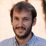
About the Presenter: Roarke Horstmeyer from Duke University
 Roarke Horstmeyer is an assistant professor of Biomedical Engineering, Electrical and Computer Engineering, and Physics at Duke University. He develops microscopes, cameras and computer algorithms for a wide range of applications, from forming large-area, high-resolution 3D videos of freely moving organisms to detecting blood flow and brain activity deep within tissue. Before joining Duke in 2018, Dr. Horstmeyer was a visiting professor at the University of Erlangen in Germany and an Einstein International Postdoctoral Fellow at Charité Medical School in Berlin. Prior to his time in Germany, Dr. Horstmeyer earned a PhD from Caltech’s EE department (2016), an MS from the MIT Media Lab (2011), and bachelor’s degrees in Physics and Japanese from Duke in 2006.
