About
05 March 2024
New cardiovascular imaging approach provides a better view of dangerous plaques
Combining FLIM with polarization sensitive OCT could lead to new ways to prevent heart attacks and strokes
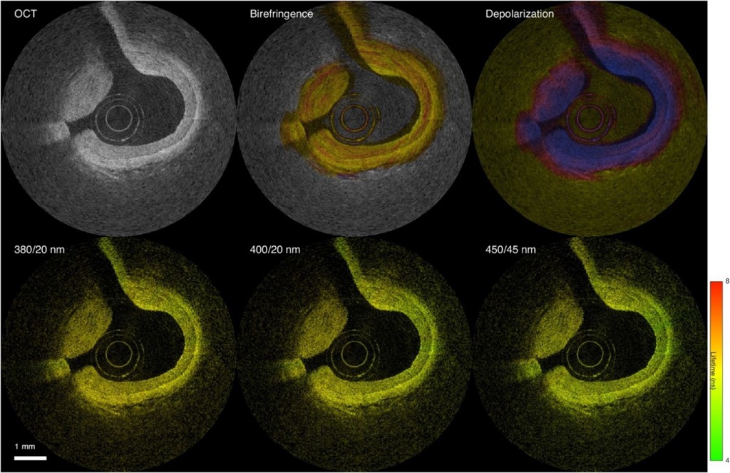
Caption: Researchers have developed a new catheter-based device that combines polarization-sensitive (PS) OCT (top) and multispectral FLIM (bottom) to provide a detailed multimodal view via intravascular imaging.
Credit: Julien Bec, University of California, Davis
WASHINGTON — Researchers have developed a new catheter-based device that combines two powerful optical techniques to image the dangerous plaques that can build up inside the arteries that supply blood to the heart. By providing new details about plaque, the device could help clinicians and researchers improve treatments for preventing heart attacks and strokes.
Atherosclerosis occurs when fats, cholesterol and other substances accumulate on the artery walls, which can cause these vessels to become thick and stiff. A heart attack or stroke may occur if an atherosclerotic plaque inside the blood vessels ruptures or parts of it break off.
“Atherosclerosis, leading to heart attacks and strokes, is the number one cause of death in Western societies — exceeding all combined cancer types — and, therefore, a major public health issue,” said research team member leader Laura Marcu from University of California, Davis. “Better clinical management made possible by advanced intravascular imaging tools will benefit patients by providing more accurate information to help cardiologists tailor treatment or by supporting the development of new therapies.”
In the Optica Publishing Group journal Biomedical Optics Express, researchers describe their new flexible device, which combines fluorescence lifetime imaging (FLIM) and polarization-sensitive optical coherence tomography (PSOCT) to capture rich information about the composition, morphology and microstructure of atherosclerotic plaques. The work was a collaborative project with Brett Bouma and Martin Villiger, experts in OCT from the Wellman Center for Photomedicine at Massachusetts General Hospital.
“With further testing and development, our device could be used for longitudinal studies where intravascular imaging is obtained from the same patients at different timepoints, providing a picture of plaque evolution or response to therapeutic interventions,” said Julien Bec, first author of the paper. “This will be very valuable to better understand disease evolution, evaluate the efficacy of new drugs and treatments and guide stenting procedures used to restore normal blood flow.”
Gaining an unprecedented view
Most of what scientists know about how atherosclerosis forms and develops over time comes from histopathology studies of postmortem coronary specimens. Although the development of imaging systems such as intravascular ultrasound and intravascular OCT has made it possible to study plaques in living patients, there is still a need for improved methods and tools to investigate and characterize atherosclerosis.
To address this need, the researchers embarked on a multi-year research project to develop and validate multispectral FLIM as an intravascular imaging modality. FLIM can provide insights into features such as the composition of the extracellular matrix (the protein scaffolding between cells), the presence of inflammation and the degree of calcification inside an artery. In earlier work, they combined FLIM with intravascular ultrasound, and in this new work they combined it with PSOCT. PSOCT provides high-resolution morphological information along with birefringence and depolarization measurements. When used together, FLIM and PSOCT provide an unprecedented amount of information on plaque morphology, microstructure and biochemical composition.
“Birefringence provides information about the plaque collagen, a key structural protein that helps with lesion stabilization, and depolarization is related to lipid content that contributes to plaque destabilization,” said Bec. “Holistically, this hybrid approach can provide the most detailed picture of plaque characteristics of all intravascular imaging modalities reported to date.”
Getting two imaging modalities into one device
The development of multimodal intravascular imaging systems compatible with coronary catheterization is technologically challenging. It requires very thin — less than 1 mm — flexible catheters that can operate in vessels with sharp twists and turns. A high imaging speed of around 100 frames/second is also necessary to limit cardiac motion artifacts and ensure proper imaging inside an artery.
To integrate FLIM and PSOCT into a single device without compromising the performance of either imaging modality, the researchers used optical components previously developed by Marcu’s lab and other research groups. Key to achieving high PSOCT performance was a newly designed rotary collimator with high light throughput and a high return loss — the ratio of power reflected back toward the light source compared to the power incident on the device. The catheter system they developed has similar dimensions and flexibility as the intravascular imaging devices that are currently in clinical use.
After testing the new system with artificial tissue to demonstrate basic functionality on well characterized samples, the researchers also showed that it could be used to measure properties of a healthy coronary artery removed from a pig. Finally, in vivo testing in swine hearts demonstrated that the hybrid catheter system’s performance was sufficient to support work toward clinical validation. These tests all showed that the FLIM-PSOCT catheter system could simultaneously acquire co-registered FLIM data over four distinct spectral bands and PSOCT backscattered intensity, birefringence and depolarization information.
Next, the researchers plan to use the intravascular imaging system to image plaques in ex vivo human coronary arteries. By comparing the optical signals acquired using the system with plaque characteristics identified by expert pathologists, they can better understand which features can be identified by FLIM-PSOCT and use this to develop prediction models. They also plan to move forward with testing in support of clinical validation of the system in patients.
Paper: J. Bec, X. Zhou, M. Villiger, J. A. Southard, B. Bouma, L. Marcu, “Dual modality intravascular catheter system combining pulse-sampling fluorescence lifetime imaging and polarization-sensitive optical coherence tomography,” Biomed. Opt. Express, Vol. 15, Issue 4, pp. 2114-2132 (2024).
DOI: doi.org/10.1364/BOE.516515
About Optica Publishing Group
Optica Publishing Group is a division of the society, Optica, Advancing Optics and Photonics Worldwide. It publishes the largest collection of peer-reviewed and most-cited content in optics and photonics, including 18 prestigious journals, the society’s flagship member magazine, and papers and videos from more than 835 conferences. With over 400,000 journal articles, conference papers and videos to search, discover and access, our publications portfolio represents the full range of research in the field from around the globe.
About Biomedical Optics Express
Biomedical Optics Express serves the biomedical optics community with rapid, open-access, peer-reviewed papers related to optics, photonics and imaging in biomedicine. The journal scope encompasses fundamental research, technology development, biomedical studies and clinical applications. It is published monthly by Optica Publishing Group and edited by Christoph Hitzenberger, Medical University of Vienna, Austria. For more information, visit Biomedical Optics Express.
Media Contact
