Some counterintuitive lessons learned from the OSA BIOMED meeting
Kyle Quinn
1. You don’t need a nice objective to achieve sub-micron optical resolution
In my lab, we are rather protective of our low magnification, high NA, long working distance microscope objectives. These objectives cost tens of thousands of dollars, but it’s hard to beat them when you want to acquire high-resolution, 10 megapixel images. However, on the last day of the BIOMED meeting, Dr. Changhuei Yang from Cal Tech demonstrated how we can acquire gigapixel images with sub-micron resolution using a 0.08 NA objective. His lab utilizes Fourier Ptychographic Microscopy, which only requires the incorporation of a simple LED array to illuminate your sample from different angles within a conventional microscope setup. Phase information is retrieved from multiple images corresponding to the illumination of different LEDs, and this algorithm enables the recovery of an image with significantly enhanced resolution. Although the approach is a little computationally intensive (~30 seconds using a GPU) and currently cannot be used for fluorescence imaging or thick samples, it’s a remarkable advancement to extend the conventional limits of microscopy.
2. Longer wavelengths do not necessarily translate to deeper imaging depths
The OSA BIOMED meeting appeals for many reasons - one of which is its smaller size, which helps facilitate one-on-one interactions with leaders in the biomedical optics field, such as Dr. Paul Campagnola. In chatting with Dr. Campagnola, I learned of his group’s recent publication in Optics Letters, which suggests longer wavelengths are not necessarily better for second harmonic generation imaging deep within tissues. Although one would think that the decreased scattering associated with longer wavelengths would enable improved SHG imaging deeper within tissues, Dr. Campagnola asserts that this is not the case because the magnitude of collagen’s first order hyperpolarizability tensor, β, rapidly decreases at these longer wavelengths. β decreases because longer wavelengths do not overlap with collagen’s absorption bands and there is a reduction in resonance-enhanced SHG that far outweighs the benefits of reduced scattering. Although it’s not what I would have expected, his computational and experimental data suggest these shorter wavelengths are better.
3. Everyone seems to be going with the flow
In a follow up to my initial post analyzing the frequency of words in previous BIOMED programs, I decided to quantify any emerging trends based on the final 2014 BIOMED program book. Not surprisingly I found that photoacoustic imaging continues to grow with a 32% increase in frequency. Interestingly, the other clear trend was the increase relative to the 2012 meeting in studies looking at blood (20.5% increase) and flow (33.8% increase). A quick survey of the program suggests no single technology is driving this increased interest in blood flow, as blood flow studies involving near-infrared spectroscopy, speckle contrast optical tomography, multi-photon microscopy, and OCT could be found.

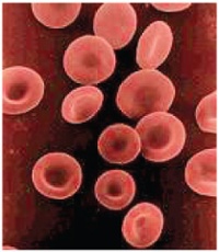
Image credit: Illustration from Anatomy & Physiology, Connexions Web site. http://cnx.org/content/col11496/1.6/, Jun 19, 2013.(CC BY 3.0)
