About
16 June 2022
AI-based image analysis automatically detects serious heart condition
New method makes it easier to detect plaque erosion using intravascular OCT images
WASHINGTON — Researchers have developed a new artificial intelligence (AI) method that can automatically detect plaque erosion in the heart’s arteries using optical coherence tomography (OCT) images. Monitoring plaque in the arteries is important because when plaque breaks apart it can block blood flow to the heart, leading to a heart attack or other serious conditions.
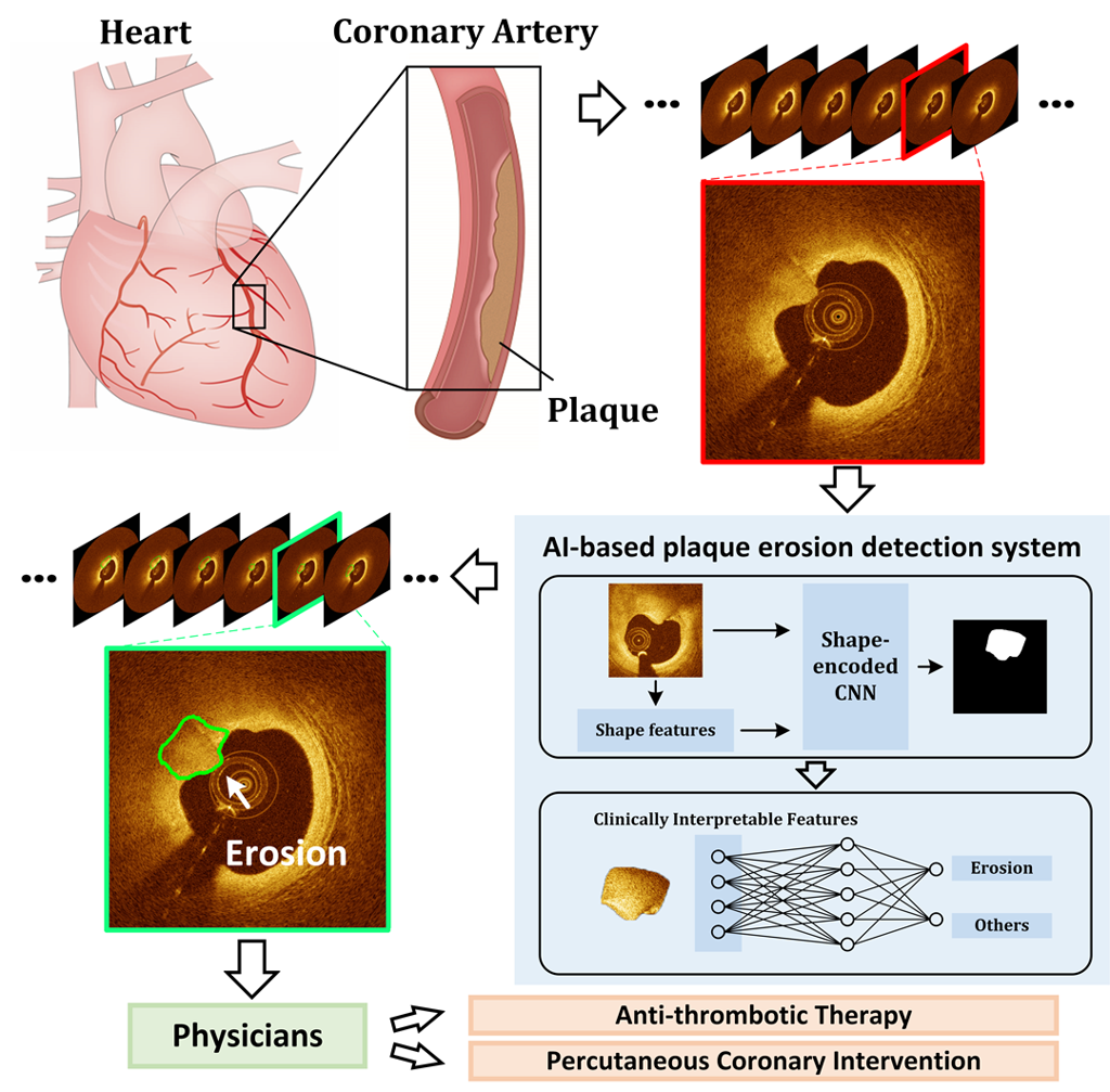
Caption: Researchers have developed a new AI method that can automatically detect plaque erosion in arteries using OCT images. This type of erosion can block blood flow to the heart, leading to a heart attack or other serious conditions.
Image Credit:Zhao Wang, University of Electronic Science and Technology of China
“If cholesterol plaque lining arteries starts to erode it can lead to a sudden reduction in blood flow to the heart known as acute coronary syndrome, which requires urgent treatment,” said research team leader Zhao Wang from the University of Electronic Science and Technology of China. “Our new method could help improve the clinical diagnosis of plaque erosion and be used to develop new treatments for patients with heart disease.”
OCT is an optical imaging method with micron-scale resolution that, when integrated with a miniaturized catheter, can be used within blood vessels to provide 3D images of the coronary arteries that supply blood to the heart. Although clinicians are increasingly using intravascular OCT to look for plaque erosion, the large amount of data produced and the complexity of visually interpreting the images has led to significant interobserver variability.
To solve this problem, Wang worked with a group of engineers from his institution and physicians led by Bo Yu from The 2nd Affiliated Hospital of Harbin Medical University to develop an objective, automatic method that uses AI to detect plaque erosion based on OCT images. They describe the new technique in the Optica Publishing Group journal Biomedical Optics Express and show that it is precise enough to potentially be used as a basis for clinical diagnosis.
“Our new AI-based method can automatically detect the presence of plaque erosion using the original OCT images without any additional input,” said Wang. “The ability to detect plaque erosion objectively and automatically will reduce the laborious manual assessment associated with diagnosis.”
Applying AI
The new method consists of two primary steps. First, an AI model known as a neural network uses the original image and two pieces of shape information to predict regions of possible plaque erosion. The initial prediction is then refined with a post-processing algorithm based on clinically interpretable features that mimic the knowledge professional physicians use to make a diagnosis.
“We had to develop a new AI model that incorporates explicit shape information, the key feature used to identify plaque erosion in OCT images,” said Wang. “The underlying intravascular OCT imaging technology is also crucial because it is currently the highest resolution imaging modality that can be used to diagnose plaque erosion in living patients.”
When OCT is used for intravascular imaging, the imaging probe is automatically pulled backward inside a catheter, producing hundreds of images for each pullback. The researchers tested their method using 16 pullbacks of 5,553 clinical OCT images with plaque erosion and 10 pullbacks of 3,224 images without plaque erosion. The automated method correctly predicted 80 percent of the plaque erosion cases with a positive predictive value of 73 percent. They also found that diagnoses based on the automated method matched well with those from three experienced physicians.
“Although further safety validation and regulatory approval are needed for stand-alone clinical use in patients, the technique could be used to facilitate diagnosis of plaque erosion,” said Wang. “This would involve physicians making a final check of the algorithm’s finding and then determining the cause of acute coronary syndrome and the best treatment strategies.”
Studying new treatments
The method could also be useful for analyzing the massive amounts of existing OCT data by eliminating the time-consuming and tedious process of manual image analysis. This could help scientists improve identification and treatment for plaque erosion. For example, a stent is often used to recover reduced blood flow in patients with acute coronary syndrome, but recent studies suggest that some medications might offer a less-invasive alternative.
“Intravascular imaging, accompanied with AI technologies, can be an extremely valuable tool for diagnosis of coronary artery disease and treatment planning,” said Wang. “In the future, this new approach could help physicians develop individualized treatment strategies for optimal management of patients with acute coronary syndrome.”
The researchers are now working to improve their new technique by better incorporating 3D information and incorporating more unlabeled data to improve the AI model’s performance. In the future, they also plan to use a larger dataset that includes a global population for training and evaluating the algorithm. They also want to explore how it might be used in various clinical situations to further demonstrate its potential utility and value.
Paper: H. Sun, C. Zhao, Y. Qin, C. Li, H. Jia, B. Yu, Z. Wang, “In Vivo Detection of Plaque Erosion by Intravascular Optical Coherence Tomography Using Artificial Intelligence,” Biomed. Opt. Express, Vol. 13, Issue 7, pp. 3922-3938 (2022)
DOI: https://doi.org/10.1364/BOE.459623
About Optica Publishing Group
Optica Publishing Group is a division of the society, Optica, Advancing Optics and Photonics Worldwide. It publishes the largest collection of peer-reviewed and most-cited content in optics and photonics, including 18 prestigious journals, the society’s flagship member magazine, and papers and videos from more than 835 conferences. With over 400,000 journal articles, conference papers and videos to search, discover and access, our publications portfolio represents the full range of research in the field from around the globe.
About Biomedical Optics Express
Biomedical Optics Express serves the biomedical optics community with rapid, open-access, peer-reviewed papers related to optics, photonics and imaging in biomedicine. The journal scope encompasses fundamental research, technology development, biomedical studies and clinical applications. It is published monthly by Optica Publishing Group and edited by Christoph Hitzenberger, Medical University of Vienna, Austria. For more information, visit Biomedical Optics Express.
Media Contact
