Pushing the limits of imaging resolution and penetration depth
Kyle Quinn
The development of labeling techniques capable of providing customizable molecular specificity has made optical microscopy a fundamental technique in the biomedical research, and the standard compound microscope remains a fixture in just about any clinic or biomedical lab. The popularity of optical microscopy was also driven by the ability to provide resolution at the cellular level that traditional clinical imaging modalities (e.g. ultrasound, x-ray CT, and MRI) simply cannot meet. The finer structural details of biological tissues were further elucidated through the development of transmission electron microscopy, which enabled unparalleled views at the scale of proteins and molecules. However, like all imaging technologies, there is a tradeoff between imaging resolution and penetration depth, and electron microscopy has extremely limited penetration depth.
Most optical techniques occupy a sweet spot with image resolution that is far superior to the traditional clinical imaging modalities and penetration depth ranging from microns to centimeters that is far better than electron microscopy. With the ability to resolve cellular and subcellular features, optical techniques offer great potential for detecting the early stages of disease or trauma and elucidating their underlying mechanisms. The development of laser scanning confocal microscopy provided depth resolution without the mechanical tissue sectioning necessary for conventional wide-field microscopy. As a result, 3D tissues could be imaged at depths of 100s of microns. Using pulsed lasers, the depth of penetration is being further pushed to 1mm and beyond through multi-photon (MP) microscopy. As a result, microscopy techniques are becoming far more amenable to non-destructive assessments in the clinic. More recently, stimulated emission depletion microscopy (STED), photoactivated localization microscopy (PALM), stochastic optical reconstruction microscopy (STORM), and other super-resolution techniques have been developed to non-destructively provide images of dynamic cellular processes at an unprecedented resolution. In Miami, at OSA BIOMED 2014, a topic section dedicated recent developments in optical microscopy techniques (BIO 3) will explore recent efforts to continue to push the limits of imaging resolution and depth.
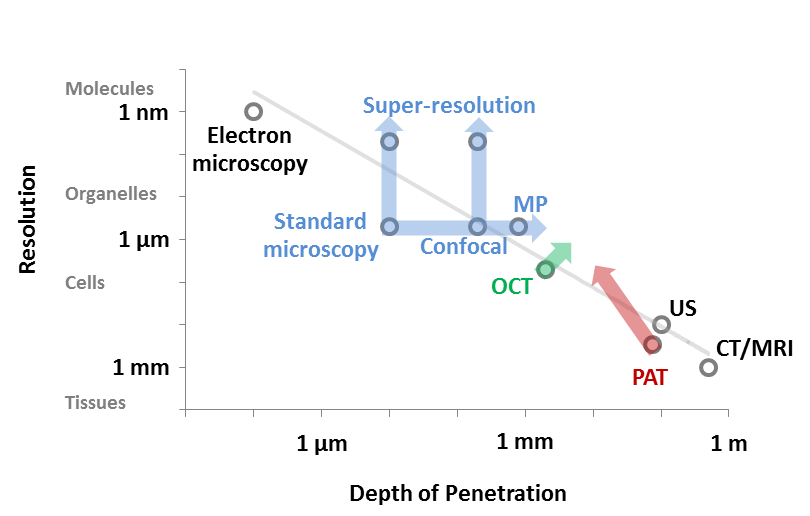
Over the last 20 years, additional optical techniques have been developed, such as optical coherence tomography (OCT) and photoacoustic tomography (PAT), which have helped to extend the range of optical imaging modalities to penetration depths that that exceed 1mm, yet still offer resolution on the order of microns to millimeters. OSA BIOMED has dedicated topic sections to both photoacoustic imaging (BIO 4), and OCT (BIO 5) as well. Collectively, biomedical optics has expanded to cover the full spectrum of length scales between the ultrastructure visible through electron microscopy to the macro-scale view of CT and MRI. I look forward to learning of the latest approaches to pushing these limits of resolution and penetration depth in Miami.
